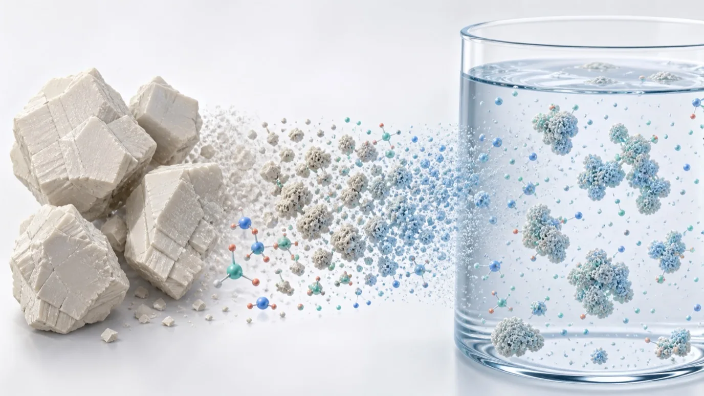
Engineering CaHA from the micrometer scale into a nano- or submicron-scale system changes the most visible specification: size. In an inorganic biomaterial, however, the same process can also alter crystallinity, surface chemistry, aggregation, and the way the material contacts cells.
A study published in August 2026 in the Journal of Biomedical Materials Research Part B: Applied Biomaterials started with commercial CaHA of approximately 10 μm and used nano spray drying to prepare a lactose-stabilized nanostructured system. The investigators then compared its physicochemical properties, fibroblast responses, and calcium delivery into ex vivo porcine skin.
The central materials question is not whether a smaller particle produced a stronger response. It is whether a system in which size, processing history, dispersion medium, and structural order all changed can still be defined by one number. In this case, nanoCaHA is not a complete material description.
What Does 181 nm Actually Describe?
The reported value of 181.06 ± 23.83 nm was obtained by dynamic light scattering (DLS) after dispersing the sample in ethanol. It is a hydrodynamic diameter under defined test conditions, not a direct geometric measurement of the dry particles.
When the same material was dispersed in ultrapure water, its hydrodynamic diameter increased to 3061.44 ± 725.61 nm, or about 3.06 μm. The zeta potential in water was +11.75 ± 3.63 mV. This modest positive value describes surface charge under that condition; it does not, by itself, demonstrate colloidal stability. The much larger hydrodynamic size in water makes particle interaction and aggregation part of the material description.
| Test condition | Reported result | Interpretation |
|---|---|---|
| DLS in ethanol | 181.06 ± 23.83 nm; PDI 0.23 ± 0.17 | Submicron hydrodynamic size in this dispersion medium |
| DLS in ultrapure water | 3061.44 ± 725.61 nm | Greater particle interaction or aggregation after the medium changed |
| Zeta potential in water | +11.75 ± 3.63 mV | A conditional surface-charge result, not a substitute for dispersion-stability testing |
This follows the same measurement principle discussed in our article on why CaHA D50 and SEM observations do not directly match: establish what was measured before interpreting the number.
Nano Spray Drying Changed More Than Size
The study did not mechanically reduce a 10 μm CaHA particle while holding every other variable constant. The starting CaHA was dispersed in water adjusted to pH 3 with acetic acid, combined with 10% w/w lactose, and processed by nano spray drying.
The resulting system therefore differed from the starting material in several ways at once: particle dimensions and morphology changed, lactose contributed to an organic-inorganic interface, the acidic process influenced surface state, and both structural order and dispersion behavior were altered. The biological differences cannot be assigned exclusively to smaller size.
The authors included a lactose-only control and reported that nanostructured CaHA prepared without lactose retained collagen-related activity. The latter result was described as preliminary and the data were not shown. It reduces the likelihood that lactose alone explains the entire response, but it does not isolate the contributions of size, amorphization, surface state, and processing history.
The XRD Pattern Shows a Different Material State
Commercial CaHA showed identifiable crystalline diffraction peaks. After processing, those characteristic peaks were replaced by a broad amorphous halo centered near 2θ = 30°. FTIR also showed broader phosphate bands and absorptions consistent with Type B carbonated hydroxyapatite.
The result should not be summarized as “XRD still confirmed the same CaHA, only at a smaller size.” The study continued to describe a CaHA-derived nanostructured system, but the combined XRD and FTIR evidence indicates a low-order, carbonated material containing a lactose-associated interface.
Similarity to selected features of biological apatite is not a performance endpoint. Carbonate substitution, amorphous content, ion release, and aqueous stability remain separate questions and should not be replaced by the word biomimetic.
The Collagen Findings Are Not a Size-Only Effect
The study used human dermal fibroblasts and, in selected experiments, a Transwell co-culture with peripheral blood mononuclear cells (PBMCs) from three independent donors.
Under co-culture conditions, COL1A1 mRNA increased by an average of approximately fivefold and COL3A1 mRNA by approximately 25-fold relative to the vehicle control. Some endpoints were also higher than those observed with the commercial CaHA formulation used as a comparator. These are transcriptional results; they do not establish that type I and type III collagen proteins were deposited in the same proportions. Sirius Red was used separately to assess total collagen and cannot resolve the protein contribution of each subtype.
The paper also reported sustained collagen production five days after treatment withdrawal. That extended observation used an adjusted design involving PBMC-conditioned medium rather than simply continuing the original co-culture unchanged.
SEM and EDS showed calcium- and phosphorus-rich deposits associated with fibroblast surfaces and the surrounding matrix after washout. Their intracellular or extracellular location was not established, and persistence was not shown to be the sole cause of the prolonged collagen signal.
More Calcium in Skin Does Not Prove Intact-Particle Permeation
In the ex vivo porcine-skin diffusion experiment, total calcium measured 34.53 ± 6.64 μg/cm² after nanoCaHA exposure, compared with 23.43 ± 3.96 μg/cm² in the control. The assay used tissue digestion followed by flame atomic absorption spectrometry, so it measured total calcium recovered from the skin sample.
The method did not specifically track intact particles. It therefore cannot distinguish intact nanoCaHA from dissolved calcium or calcium-containing material retained at different skin depths. It does not independently prove that intact 181 nm particles crossed the complete skin barrier, nor does it establish the safety or dermal efficacy of a topical formulation in humans.
The authors proposed superficial and transdermal development opportunities while also stating that irritation, sensitization, and clinical studies of the complete formulation remain necessary before human use. The present evidence is limited to cell experiments and an ex vivo skin model.
How Should nanoCaHA Be Described?
At the nanoscale, an average size is not enough. A useful material file should also identify:
- whether size came from DLS, SEM, TEM, or another method;
- the dispersion medium, concentration, sonication, and dispersant conditions;
- hydrodynamic size, PDI, zeta potential, and repeatability;
- dry-state morphology and aqueous aggregation;
- crystalline and amorphous content, carbonate substitution, and surface chemistry;
- changes in size and interface after contact with proteins or biological fluids.
From a specification perspective, nanoCaHA is not a self-sufficient material identity. The 181 nm result in ethanol, the approximately 3 μm result in water, the amorphous halo, Type B carbonation, and the lactose-associated interface describe different aspects of the same experimental system. They must be interpreted together.
Conflict-of-interest disclosure: the paper states that four authors are inventors on a patent directed to the invention and that two of those four authors are co-founders of AMA (Ativos Modernos Atomizados). This article is a public materials-science review. The reported findings come from cell and ex vivo porcine-skin experiments and do not establish human clinical safety, effectiveness, or the performance of Nanjing Junzhuo's current CaHA/HAp products.
Reference
- Bispo ECI, de Barcelos SM, Contarato JLA, et al. Nanostructured and Functionalized Calcium Hydroxyapatite Enhances Collagen Production by Human Dermal Fibroblasts Compared With Microstructured Counterparts In Vitro. Journal of Biomedical Materials Research Part B: Applied Biomaterials. 2026;114(8):e70144. DOI: 10.1002/jbm.b.70144.