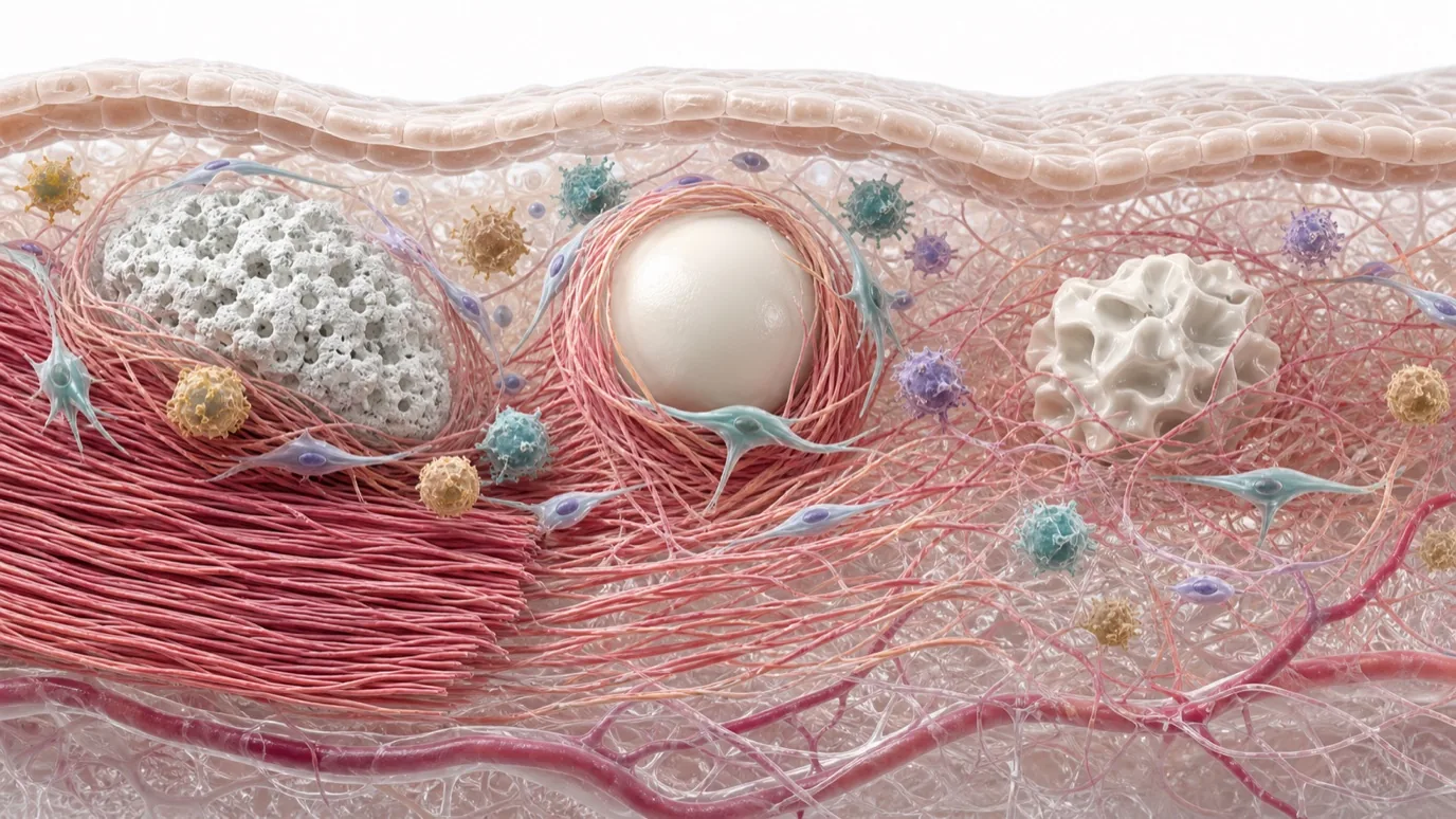
“More collagen” is one of the most familiar endpoints in biostimulatory-material research. Stained area, COL1A1 expression, and total collagen content can all be informative, but they do not answer the same question.
Two materials can produce similar total-collagen values without producing the same tissue result. Collagen subtype, fiber organization, the surrounding immune state, and the observation time can change the biological meaning of the number.
In August 2026, Ye et al. published a peer-reviewed accepted manuscript in npj Regenerative Medicine comparing CaHA, PCL, spherical PLLA (PLLA-Sp), and irregular PLLA particles (PLLA-Ir). The study placed material characterization, fibroblast and macrophage responses, collagen subtypes, and rat intradermal tissue findings in one multiscale framework. The accepted manuscript is citable and has a permanent DOI, but copyediting and presentation may change before the Version of Record.
The Highest Total Collagen Did Not Mean the Same Tissue Outcome
PLLA-Sp produced the greatest total collagen deposition in the study. If evaluation stopped there, it could easily be described as the strongest collagen stimulus. Further analysis showed predominant type III collagen and persistent mixed inflammation in the PLLA-Sp group. CaHA did not produce the highest total collagen, but it was associated with denser, more organized collagen bundles, a higher type I/III collagen ratio, and a higher CD206/CD68 ratio.
These findings do not create a simple ranking. They show that the amount of collagen and the organization of the resulting matrix are separate evaluation dimensions.
| Evaluation dimension | Question answered | What it cannot establish alone |
|---|---|---|
| Total collagen | Overall change in collagen deposition within the assay | Subtype, organization, or long-term matrix maturation |
| Type I/III ratio | Relative composition at a defined time point | That a higher type I fraction necessarily produces a better clinical result |
| Fiber organization | Density and arrangement of collagen bundles | Tissue mechanics or long-term stability |
| Immune markers | One window into local immune-cell state | The complete inflammatory trajectory or human safety |
Type I and Type III Collagen Need a Time Context
Type I is the predominant structural collagen in adult skin. Type III is also a normal tissue component and is often associated with early repair and matrix remodeling. Reducing type I to “good” and type III to “bad” does not reflect the biology of tissue repair.
An increase in type III collagen during early remodeling may not have the same meaning as persistent type III predominance in a prolonged inflammatory environment. A stronger assessment records total amount, type I/III balance, fiber organization, and change over time without assigning a fixed value to either subtype.
The phrase collagen quality also needs definition. If used, it should be unpacked into subtype, orientation, bundle density, matrix maturation, or the surrounding inflammatory state rather than treated as one standardized endpoint.
Macrophages Are Part of the Remodeling Process
The study did not examine fibroblasts alone. It also evaluated RAW264.7 macrophages, cytokines, and polarization markers in mono- and co-culture. All tested materials promoted some degree of M2-like polarization, but the local immune environments were not identical.
CaHA showed a higher CD206/CD68 ratio. CD68 is commonly used to identify macrophage populations, while CD206 is associated with selected repair-related phenotypes. The ratio can describe an immune tendency at a defined time point; it is not a composite score of safety or regenerative performance.
The familiar equation “M1 is bad and M2 is good” is also too simple. Tissue macrophages occupy a changing continuum shaped by material surfaces, degradation, cytokines, and time. The more useful questions are how inflammation develops, how long it persists, and whether it progresses toward organized remodeling.
Surface Properties and YAP Were Associated, Not Isolated as a Single Cause
A strength of the study was that it did not compare CaHA, PCL, and PLLA as chemical labels alone. It also examined SEM and FTIR features, zeta potential, AFM roughness and stiffness, tapped density, aggregation, and rheology after incorporation into HA matrices.
Among the experimental materials, CaHA had the highest surface roughness, stiffness, and tapped density, together with a porous nanosurface and relatively low aggregation. The CaHA group also showed stronger fibroblast cytoskeletal organization, YAP nuclear translocation, and type I collagen-related responses.
YAP is an important regulator of mechanotransduction, and its localization can reflect cellular responses to mechanical cues. However, chemistry, topography, stiffness, aggregation, and other variables changed together in this comparison. There was no control in which roughness alone was altered while every other factor remained constant. The study therefore supports an association between material features and cellular responses, not proof that roughness caused a particular collagen outcome through YAP.
The Study Does Not Rank Entire Material Classes
The work combined cell experiments with a rat intradermal model. It was not a randomized clinical comparison of finished products. CaHA, PCL, PLLA-Sp, and PLLA-Ir refer to the specific experimental materials used in the paper, not every commercial product in those categories.
The acknowledgements state that experimental materials were provided by four companies, while the authors declared no competing interests. Source disclosure helps define the study conditions but does not justify extending one material result to an entire clinical category.
More organized bundles, a higher type I/III ratio, or a higher CD206/CD68 ratio are not validated surrogate endpoints for clinical effectiveness. Tissue appearance, mechanical function, durability, and human outcomes require their own study designs.
A More Complete Framework Combines Time and Tissue Outcome
Evaluation of a collagen-stimulating material can be organized across four levels:
- Material: chemistry, dimensions, surface, stiffness, aggregation, and finished-formulation state;
- Cell: fibroblast adhesion, cytoskeleton, mechanotransduction, and collagen-related expression;
- Immune: macrophage state, cytokines, and their change over time;
- Tissue: total collagen, subtype balance, fiber organization, inflammatory infiltrate, and later remodeling.
These levels can support mechanistic hypotheses, but they are not interchangeable. Gene expression does not replace collagen-protein deposition. A histology image does not independently establish long-term mechanical improvement. Organized collagen in an animal model is not a human clinical claim.
The logic is similar to our discussion of the degradation microenvironment: Day 0 material specifications are the starting point, while tissue outcomes need a time axis. A mature assessment asks not only how much collagen increased, but which collagen, how it was organized, what immune state accompanied it, and whether those changes persisted.
This article is a public literature and materials-methodology review. The source is currently a citable peer-reviewed accepted manuscript and may undergo editorial changes before its Version of Record. The findings concern specific CaHA, PCL, and PLLA experimental materials in cell and rat studies. They do not rank material classes or commercial products clinically and do not establish the performance of Nanjing Junzhuo's current CaHA/HAp products.
Reference
- Ye L, Wang L, Jin P, et al. Multiscale evaluation of collagen-stimulating materials: linking physicochemical properties to dermal regeneration outcomes. npj Regenerative Medicine. Published online August 14, 2026. DOI: 10.1038/s41536-026-00498-3.