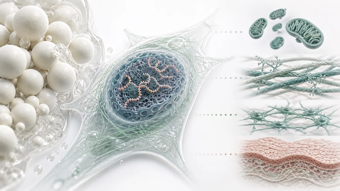
Mechanistic studies of CaHA have often focused on collagen expression and extracellular-matrix remodeling. An in vitro study by Haddad et al., published in Life in August 2026, extended that view to SIRT1, FOXO3, SIRT3, and genes associated with mitochondrial stress and energy metabolism.
The findings broaden the research question, but they do not show that CaHA “activates an anti-aging pathway” or restores a youthful cellular state. The study measured transcription. Protein abundance, pathway activity, mitochondrial function, tissue remodeling, and clinically meaningful skin changes remain separate levels of evidence.
First, Understand What the Aging-Like Model Represents
The researchers used primary human dermal fibroblasts and exposed them to 10 μM EX-527, a SIRT1 inhibitor, for 48 hours. EX-527 remained present during the subsequent 72-hour CaHA treatment period. FOXO3, SIRT1, and SIRT3 expression decreased after inhibition, while representative SA-β-gal staining increased. The authors therefore described the system as a SIRT1-inhibition-associated aging-like state.
This model is useful for observing cellular responses when SIRT1 is suppressed, but it is not a complete model of human skin aging. Natural and photoinduced aging involve ultraviolet exposure, oxidative stress, DNA damage, extracellular-matrix mechanics, chronic inflammation, and long-term metabolic changes. The paper also notes that p16, p21, DNA-damage markers, and broader SASP profiling were not used to validate strict cellular senescence.
| Study element | Condition used | Interpretive limit |
|---|---|---|
| Cell model | Primary human dermal fibroblasts | Two-dimensional culture, not intact skin |
| Aging-like induction | 10 μM EX-527 for 48 hours and maintained thereafter | Models a SIRT1-inhibited state, not natural aging |
| Material exposure | Three commercial CaHA formulations for 72 hours | One concentration and one time point |
| Primary method | RT-qPCR | Measures mRNA, not protein or cellular function |
Several Gene Groups Showed Transcriptional Responses
After 72 hours under the same experimental conditions, all three commercial CaHA formulations increased COL1A1 and Ki67 expression. ELN, cytokine-related genes, and homeostasis-associated genes showed formulation-dependent magnitudes and directions.
SIRT1 expression increased by about 37.4% with sample R and 35.4% with sample S, compared with 5.9% for sample D, which did not reach significance. Sample S increased FOXO3 by approximately 41.4% and SIRT3 by 20.0%. PGC-1α, SOD2, and PINK1 increased by about 92.5%, 35.2%, and 38.9%, respectively.
These values support a restrained conclusion: in this in vitro system, CaHA formulation exposure was associated with differential transcriptional responses involving extracellular matrix, cell-cycle activity, inflammatory regulation, and mitochondrial stress. They generate mechanistic hypotheses; they do not complete mechanistic validation.
mRNA Is Only the First Layer of Evidence
RT-qPCR shows how much of a gene transcript is present. Functional pathway activity also depends on protein abundance, post-translational modification, cellular localization, enzyme activity, and downstream cell behavior.
| Evidence layer | What can be measured | Covered here? |
|---|---|---|
| Transcription | Changes in COL1A1, SIRT1, FOXO3, and other mRNAs | Yes |
| Protein and pathway activity | Protein abundance, enzyme activity, FOXO3 acetylation and localization | No |
| Cell function | ROS, ATP, respiration, membrane potential, proliferation, and migration | No |
| Tissue outcome | Collagen bundles, elastic fibers, dermal structure, and long-term remodeling | No |
| Clinical outcome | Skin texture, elasticity, wrinkles, and durability | No |
A separate 2026 UVA-photoaging study by Ma et al. illustrates the distinction. It measured SIRT1-mediated FOXO3a deacetylation and stabilization, then used FOXO3a knockdown to test whether the protective effects disappeared. Intervention and blocking experiments of this kind provide stronger causal evidence. The CaHA findings are more accurately described as changes in SIRT1/FOXO3-related transcription.
Mitochondrial Markers and Ki67 Need Functional Validation
PGC-1α, SOD2, and PINK1 are associated with mitochondrial biogenesis, antioxidant defense, and quality control. Higher expression may indicate an adaptive response, but it may also reflect compensation for cellular stress. Without ROS, ATP, oxygen-consumption, membrane-potential, or mitochondrial-morphology data, the results cannot establish improved mitochondrial function.
The same caution applies to Ki67. The assay measured Ki67 mRNA rather than cell counts, cell-cycle distribution, or long-term replicative capacity. Upregulation can indicate cell-cycle-related transcriptional activity, but it does not demonstrate reversal of cellular senescence.
Terms such as “anti-aging genes” or “longevity genes” also compress complex biology into a marketing label. SIRT1, FOXO3, and SIRT3 participate in several homeostatic processes, and their effects depend on cell type, timing, and stress context. “Aging-associated” or “cellular-homeostasis-related” is more consistent with the evidence.
Formulation Differences Do Not Reveal the Material Cause
The three commercial CaHA formulations produced different transcriptional patterns. This means complete CaHA formulations should not automatically be assumed to elicit identical cellular responses. It does not identify which material variable produced the difference.
The study did not pair the biological results with direct comparisons of particle-size distribution, microsphere morphology, specific surface area, CaHA concentration, carrier composition, zeta potential, or rheology. The observed differences could relate to microspheres, carrier properties, concentration, manufacturing, or several variables acting together. They cannot be used to rank clinical products or to establish superiority of a single CaHA raw-material parameter.
Ilikia Brasil funded the laboratory testing and article-processing charge. The author group included an employee, scientific consultants, and participants in company-supported educational activities. The paper states that the experiments, data generation, and statistics were conducted by an independent laboratory. These relationships do not invalidate the study, but they remain relevant context when interpreting a product comparison.
A Stronger Mechanistic Program Requires Matched Evidence
For materials development, the next step is not simply a larger gene panel. Biological endpoints should be paired with characterization of the same material batches: particle-size distribution, microsphere morphology and surface state, phase and crystallinity, specific surface area, carrier composition, formulation rheology, and dispersion state. Dose and time series are also needed.
Biological validation should then move from transcript to protein, from association to pathway blocking, and from markers to function. ROS, ATP, membrane potential, oxygen consumption, cell-cycle analysis, and three-dimensional skin models can test whether the transcriptional signals translate into measurable cellular behavior.
At the upstream-supply level, CaHA microsphere size distribution, morphology, phase purity, and batch consistency provide a traceable starting point for downstream mechanistic research. They do not replace functional or clinical validation of a complete formulation. The deeper a study moves into molecular pathways, the more clearly the material identity must be documented.
The most defensible conclusion from this 2026 study is not that CaHA makes cells younger. It identified transcriptional signals worth following. Gene expression is one layer of mechanistic evidence, not the endpoint.
This article interprets publicly available mechanistic and materials research. The findings discussed here come from an EX-527-induced aging-like, two-dimensional human dermal fibroblast model and a 72-hour RT-qPCR assessment. They do not establish human skin rejuvenation, clinical efficacy, or superiority of a specific CaHA product. In vitro transcriptional differences among commercial formulations should not be used for efficacy ranking without matched physicochemical characterization and further validation.
References
- Haddad A, Issa MCA, Tonon L, et al. Differential Modulation of Extracellular Matrix- and Longevity-Related Gene Expression by Calcium Hydroxylapatite Formulations. Life. 2026;16(9):1400. DOI: 10.3390/life16091400.
- Ma W, Jiang M, Zhang J, et al. SIRT1 protects against UVA-induced photoaging by suppressing oxidative stress and FOXO3a acetylation in human dermal fibroblasts. Journal of Photochemistry and Photobiology B: Biology. 2026;279:113451. DOI: 10.1016/j.jphotobiol.2026.113451.