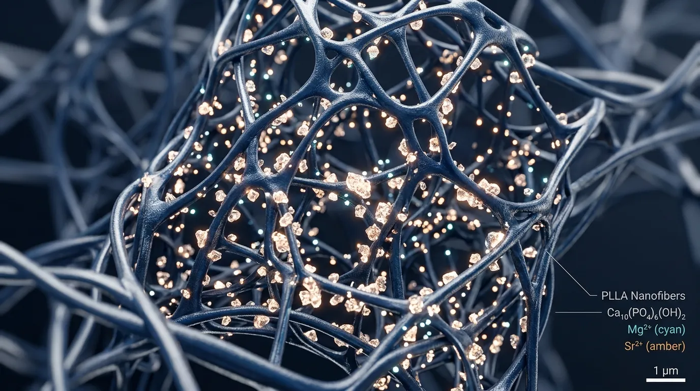
A 2026 study placed four calcium phosphate materials into the same electrospun PLLA fiber membrane. Cell proliferation was similar across groups, but mineralization was not: under osteogenic culture conditions, the magnesium/strontium co-substituted group showed the strongest mineralization.
The researchers used human periodontal ligament stem cells to examine cell growth, mineral deposits formed around the cells, and selected osteogenic genes. This was an in vitro materials study, with no animal or clinical data.
The Study Compared Four Inorganic Materials
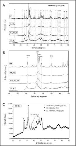
The four materials were HAp, magnesium-substituted HAp, a strontium-modified calcium phosphate, and magnesium/strontium-co-substituted HAp.
One distinction matters. The strontium-only sample was not simply strontium-substituted HAp. X-ray diffraction showed several strontium-bearing calcium phosphate phases, and the paper designated this material CP_Sr. Similar material names do not necessarily mean the same crystal phase.
The Five Membranes Had Similar Fiber Morphology
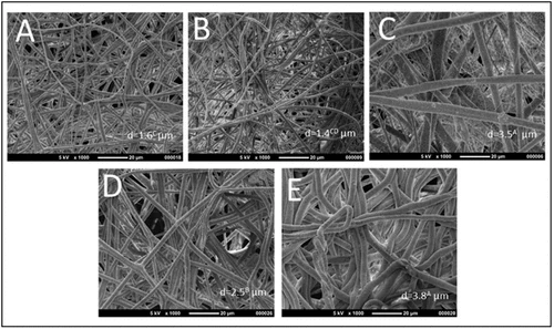
All four inorganic powders consisted of needle-shaped nanocrystals. Once incorporated into PLLA, the membranes remained randomly arranged three-dimensional fiber networks with a mean fiber diameter of about 2 μm. Fiber morphology and deposition did not differ significantly among groups.
The later cell results are therefore more plausibly linked to the composition of the inorganic phase than to visibly thicker or thinner fibers.
Cell Growth Was Similar, but Mineralization Differed
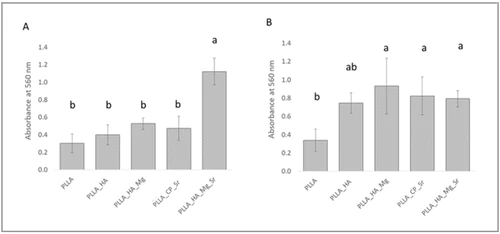
At days 3 and 7, cell proliferation did not differ significantly among groups. Cells adhered to and spread across every fiber surface. The findings do not support a simple claim that adding one ion made cells grow faster.
The main differences appeared in mineralization at day 21. In osteogenic medium, the magnesium/strontium-co-substituted HAp membrane showed the greatest mineral deposition around the cells; the other groups did not differ clearly from the PLLA control. In culture without added osteogenic induction components, the magnesium-substituted, strontium-containing calcium phosphate, and co-substituted groups all showed greater mineralization than the PLLA control.
| Readout | Result reported in the paper | How to read it |
|---|---|---|
| Cell proliferation at days 3 and 7 | No statistically significant group differences | No single ion was shown to make cells grow faster |
| Mineralization at day 21: osteogenic medium | The co-substituted group was highest | This is an in vitro result under a specific culture condition |
| Mineralization at day 21: without added induction components | Magnesium-substituted, strontium-containing, and co-substituted groups were above the PLLA control | Inorganic-phase composition affected mineral deposition |
| Gene expression | Most markers were not clearly different; OPN was higher in some groups | No universal “best material” emerged across all readouts |
Gene-Expression Results Were Mixed
The gene data did not produce the same clear ranking. Most markers did not differ substantially among groups, while only some materials showed higher osteopontin expression than the PLLA control. Stronger mineralization therefore did not mean that every osteogenic marker increased at the same time.
What Can Be Concluded So Far?
In this PLLA composite-membrane system, the way magnesium and strontium were introduced changed phase composition and was associated with different mineralization results. The co-substituted group showed the strongest mineralization in osteogenic culture conditions, but the different readouts did not identify one universal best material.
These are cell-culture findings. They do not answer how the membranes would degrade, retain mechanical properties, or form bone in animal defects or people. For composite membranes, the final crystal phases and how particles are incorporated into the fibers also matter.
This article is a public literature note. It discusses material and in vitro cell-study findings for magnesium- and strontium-modified calcium-phosphate phases in PLLA composite membranes. It does not provide medical-product recommendations, clinical indications, or treatment advice.
References
- Rodrigues MAR, Guedes HO, Galloro MB, et al. Membranes for Bone Engineering Enriched with Magnesium- and Strontium-Substituted Hydroxyapatite. ACS Omega. 2026. DOI: 10.1021/acsomega.6c01548.
- Geng Z, Cui Z, Li Z, et al. Synthesis, characterization and the formation mechanism of magnesium- and strontium-substituted hydroxyapatite. Journal of Materials Chemistry B. 2015;3:3738-3746. DOI: 10.1039/C4TB02148G.