Diluting a CaHA formulation changes particle concentration and material distribution. When the added medium contains growth factors, the biochemical environment around the cells also changes. Material dispersion, cell proliferation, and collagen-related gene expression therefore need to be evaluated as separate outcomes.
In an in vitro study first published in the Journal of Cosmetic Dermatology on October 25, 2026, Eduardo Anitua, Roberto Tierno, and Mohammad Hamdan Alkhraisat examined early responses of primary human dermal fibroblasts to undiluted Radiesse and formulations diluted with plasma rich in growth factors (PRGF). Adhesion- and proliferation-related readouts increased, while collagen-related gene expression did not follow the same pattern.
The CMC Carrier and the PRGF Diluent Have Different Roles
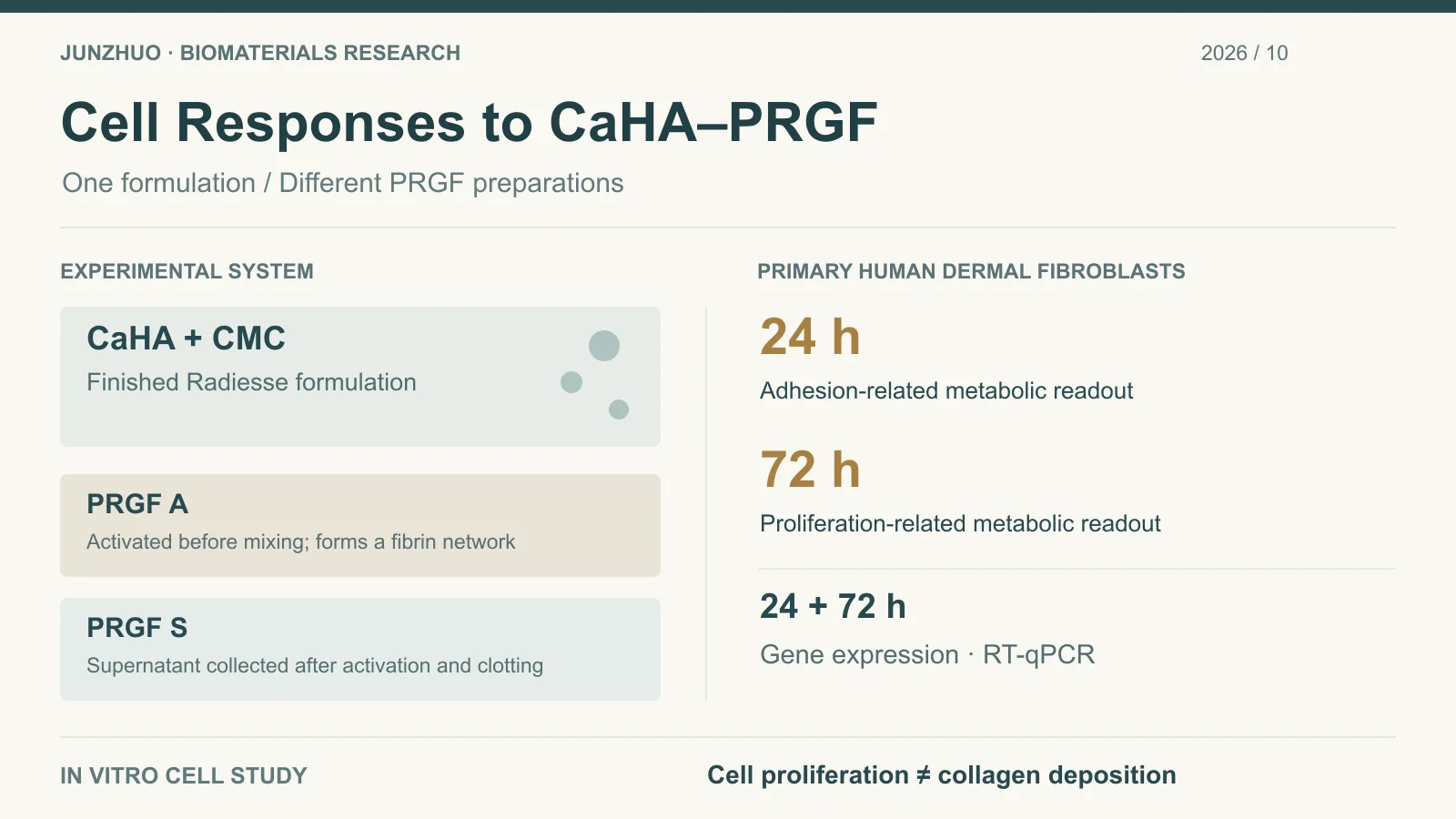
The study used the finished Radiesse formulation, with synthetic calcium hydroxylapatite (CaHA) microspheres suspended in a carboxymethylcellulose (CMC) gel carrier. PRGF was added as a diluent. Keeping these components distinct helps avoid attributing the response to the complete formulation solely to the CaHA particles.
The PRGF was a leukocyte-poor autologous platelet preparation obtained from three healthy volunteers. The researchers compared activated PRGF that was mixed with the formulation and allowed to clot (PRGF A) with supernatant collected after activation and clotting (PRGF S). The former can form a fibrin network; the latter primarily supplies components released into the liquid phase.
These preparations differ in structure and conditions for mediator release and should not be treated as one interchangeable PRGF diluent. Multiple formulation-to-PRGF ratios were evaluated alongside undiluted formulation, PRGF-only treatments, and cell-culture controls. These were experimental conditions, rather than instructions for preparing injections.
Adhesion and Proliferation Readouts Depend on the Assay
Adhesion was evaluated at 24 hours by removing non-adherent cells, washing, and using the WST-1 metabolic activity assay as an indirect readout. Proliferation was assessed at 72 hours using the same type of assay. The tests addressed different processes; they were not direct cell counts performed on every group at both time points.
PRGF mixtures generally produced higher adhesion- and proliferation-related readouts than undiluted Radiesse, with the magnitude depending on the ratio and PRGF preparation. Fibrin formed by PRGF A may contribute to adhesion, whereas PRGF S provides a different soluble-factor environment. SEM showed fibrin structures around CaHA microspheres in the PRGF A mixtures.
PRGF-only treatments also produced high readouts. A mixture outperforming undiluted formulation therefore does not, by itself, establish synergy between CaHA and PRGF. PRGF activity, diluted particle concentration, and the cell–material contact environment may all contribute.
Collagen Genes, FGF2, and MMP1 Followed Different Time Courses
RT-qPCR was used at 24 and 72 hours to examine six genes: COL1A1, COL3A1, FGF2, MMP1, TIMP1, and CTGF. The table summarizes transcriptional findings, which cannot be converted directly into protein abundance or collagen deposition.
| Marker | Main reported finding | Conditions needed for interpretation |
|---|---|---|
| COL1A1, COL3A1 | Expression was lower than with undiluted formulation under most PRGF dilution conditions | Some low-dilution conditions at 24 hours were exceptions; responses depended on ratio and time |
| FGF2 | Expression increased in PRGF-related conditions at 72 hours | Higher transcript levels do not establish increased FGF2 protein release |
| MMP1, TIMP1 | PRGF S induced an earlier MMP1 increase; TIMP1 varied over time | Transcripts do not determine collagenase activity or net matrix degradation |
| CTGF | The Results section reported lower expression with PRGF A at 72 hours than with undiluted formulation | Interpretation must retain preparation and time; neither unchanged expression nor an antifibrotic effect is a blanket conclusion |
Higher proliferation-related readouts and sustained collagen-gene upregulation are different outcomes. The authors interpreted the overall pattern as transcriptional regulation associated with early matrix turnover. Protein deposition of the investigated matrix markers was not quantified, so the direction of net collagen production or degradation remains unresolved.
The Missing Saline-Diluted Control Limits Comparisons Between Diluents
The paper explicitly notes that it did not include a normal saline-diluted Radiesse control. Comparing PRGF mixtures with undiluted formulation changes biological supplementation and physical dilution together. It cannot directly determine whether PRGF produces a different cellular response from saline at the same CaHA dose.
PRGF came from three donors, and observations were limited to 24 and 72 hours. The cell model also cannot reproduce the immune response, vascular environment, or long-term material changes in tissue. Short-term CTGF or MMP1 expression does not determine eventual fibrosis, skin firmness, or filler longevity.
The authors disclosed that all three work at BTI Biotechnology Institute, which researches PRGF-Endoret technology; Eduardo Anitua is its Scientific Director. This relationship should remain part of the study context, with independent research needed to assess reproducibility.
Specific Questions for Further CaHA Formulation Research
Further experiments could match CaHA dose and add a saline-diluted control, retaining comparisons with PRGF-only treatments and the different PRGF preparations. Separate measurements of cell number and metabolic activity, followed by collagen-protein quantification, deposition analysis, and MMP activity assays, would help determine whether increased cellular growth accompanies matrix production or degradation. These are proposed evaluations addressing the evidence gaps, rather than completed experiments in this study.
Material characterization should document CaHA particle size distribution, morphology, and phase composition, along with particle dispersion after mixing with CMC and PRGF. A network-forming PRGF A preparation and a predominantly liquid PRGF S preparation may alter cell–particle contact differently. The earlier article on CaHA/CMC dilution and material evaluation discusses physical dilution and formulation behavior; this study adds early cellular evidence for a biologically active medium.
The supported finding is that adding PRGF changed cellular readouts and gene expression in this finished CaHA formulation. Safety, efficacy, and long-term tissue remodeling after clinical mixing require separate investigation; this in vitro experiment does not establish a recommendation for injection.
Original Research
Anitua E, Tierno R, Alkhraisat MH. Dilution-Driven Regulation of Biostimulatory Signaling in Calcium Hydroxylapatite–PRGF Systems: Implications for Early Dermal Remodeling. Journal of Cosmetic Dermatology. 2026;25(11):e71257. First published October 25, 2026. DOI: 10.1111/jocd.71257.