Biphasic calcium phosphate (BCP), comprising hydroxyapatite (HAp) and β-tricalcium phosphate (β-TCP), is often designed and compared by its phase ratio. A higher fraction of one phase, however, does not guarantee a better cellular response. Cells encounter the surface left by scaffold fabrication and thermal processing, so surface chemistry and pore structure also need to be assessed.
A fish-bone-derived BCP scaffold study by Mingmuang and colleagues in the Journal of Alloys and Compounds associated differences in cellular performance with magnesium chemical state and surface pores. For HAp-based biphasic materials, this raises a practical question: which characteristics should be checked alongside XRD phase composition before interpreting differences in cell attachment and proliferation?
Two Scaffold Sources Differed in More Than Phase Ratio
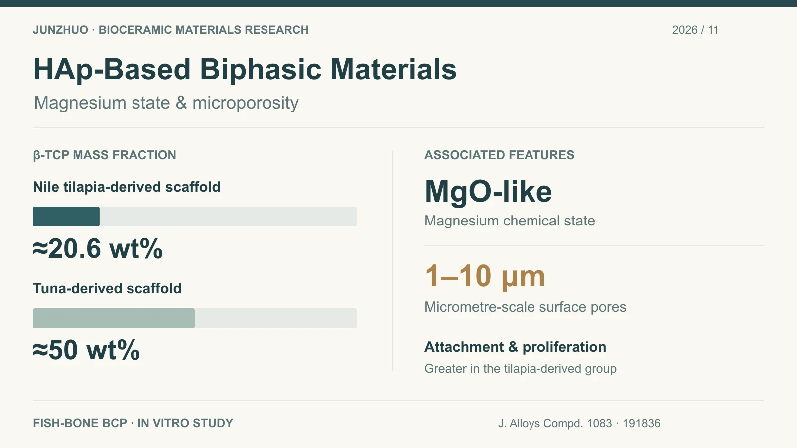
The study compared thermally processed Nile tilapia bone powder with tuna bone powder supplied by a fish-processing company, using each to prepare porous scaffolds. Both contained HAp and β-TCP. The public abstract reports the following results.
| Comparison | Nile tilapia bone-derived scaffold | Tuna bone-derived scaffold |
|---|---|---|
| β-TCP mass fraction | Approximately 20.6 wt% | Approximately 50 wt%; HAp/β-TCP close to 1:1 |
| In vitro cellular response | Significantly greater pre-osteoblastic cell attachment and proliferation | Relatively lower; both groups showed good cytocompatibility |
The scaffold with greater attachment and proliferation had magnesium predominantly in an MgO-like chemical state and a more microporous surface, with pores of 1–10 μm. Because source and processing history also differed, this comparison cannot isolate the contribution of magnesium, pore structure, or phase ratio. Reducing β-TCP content is therefore not a general prescription for improving the cellular response.
Magnesium Content and Chemical State Answer Different Questions
Total elemental analysis measures how much magnesium a sample contains; chemical-state analysis examines its chemical environment. The abstract's term MgO-like describes a chemical state. It does not, by itself, establish a separate, pure MgO crystalline phase or show that magnesium has entered the HAp lattice.
Bulk elemental composition, near-surface chemistry, and ion release into a medium should be recorded separately. Bulk magnesium measured by ICP-OES or ICP-MS cannot substitute directly for near-surface information from XPS. Detecting magnesium also does not measure its release rate. Even samples with similar total magnesium require chemical-state comparison using a consistent analytical method.
The abstract does not provide a complete quantitative relationship among magnesium content, chemical state, and ion release, so it cannot establish which is the stronger predictor. Thermal-processing studies could compare phases and magnesium environments before and after treatment, then measure release under specified medium, solid-to-liquid ratio, and sampling-time conditions.
Interpret the 1–10 μm Pores at the Micrometre Scale
The paper describes a more microporous surface with pores of 1–10 μm. Here, microporous is used in the scaffold-materials sense of micrometre-scale pores. In IUPAC physisorption terminology, micropores usually have widths no greater than approximately 2 nm. These usages should not be conflated.
SEM with defined sampling areas and image-analysis procedures can describe micrometre-scale pore dimensions, distribution, and surface morphology. Two-dimensional images do not directly give the three-dimensional porosity or interconnectivity of an entire scaffold. BET specific surface area also cannot replace direct measurement of 1–10 μm pores.
Finished scaffolds require separate assessment of overall porosity, connectivity, and mechanical properties. Whether additional surface pores coincide with lower strength, altered processing shrinkage, or local defects needs to be tested after fabrication; powder surface area alone cannot answer these questions.
Match Each Measurement to a Specific Question
For work with hydroxyapatite raw materials and biphasic calcium phosphate materials, the complementary methods below can guide evaluation. This is an editorial assessment framework, not a list of experiments confirmed as performed in the cited study.
| Question | Possible method | Scope of interpretation |
|---|---|---|
| Which crystalline phases are present, and in what proportions? | XRD; suitable refinement when quantification is required | Describes crystalline components; report the model and uncertainty for quantitative results |
| How much Ca, P, and Mg does the bulk material contain? | ICP-OES or ICP-MS after digestion | Measures elemental totals, not chemical state directly |
| What chemical environments are present at the surface? | XPS with complementary analysis appropriate to the system | Mainly near-surface information; report preparation and peak assignments |
| Which bonding and functional-group features are present? | FTIR | Provides phosphate- and hydroxyl-related vibrational information; sampling depth depends on measurement mode |
| What are the pore morphology and accessible surface area? | SEM image analysis; gas adsorption/BET | Describes morphology and specific surface area separately; BET values are not micrometre pore diameters |
| What changes occur when the material contacts a medium? | Dissolution and ion-release tests under specified conditions | Report medium, solid-to-liquid ratio, time, and normalization |
Powder, sintered scaffold, and scaffold after immersion should be sampled separately. Raw-material results establish a starting point, while fabrication and thermal treatment may change phases, chemistry, and pores. The samples used in cell experiments need corresponding characterization in their finished form.
Which Variables Should Follow-Up Experiments Control?
To examine magnesium state, studies should control powder source, fabrication, and thermal processing as far as practicable, while measuring whether phase composition and pore structure also change. Comparisons of pore architecture should likewise check composition and surface chemistry, so that several processing-induced changes are not assigned to a single pore-size effect.
Cell comparisons also require consistent scaffold dimensions, sterilization, seeding conditions, culture periods, and assays. Attachment, proliferation, and osteogenic differentiation are distinct endpoints. The first two outcomes reported in this abstract do not directly establish bone formation or clinical repair.
The study supports including magnesium chemical state and micrometre-scale surface pores in comparisons of HAp-based biphasic materials. Controlled material and cell experiments are the next step; two samples from different sources cannot establish an optimal phase ratio.
Original Research and Method References
Mingmuang Y, Pimsawat A, Khamkhantee N, et al. Beyond phase composition: Unravelling the physicochemical origins of biological performance in fish bone-derived biphasic calcium phosphate scaffolds. Journal of Alloys and Compounds. 2026;1083:191836. DOI: 10.1016/j.jallcom.2026.191836.
Study findings are drawn from the public abstract and bibliographic record at Mahidol University; the measurement and control designs above are editorial materials-development analysis. Pore terminology follows the IUPAC pore classification; the scope of XPS follows the practical guide to planning and reporting XPS measurements.